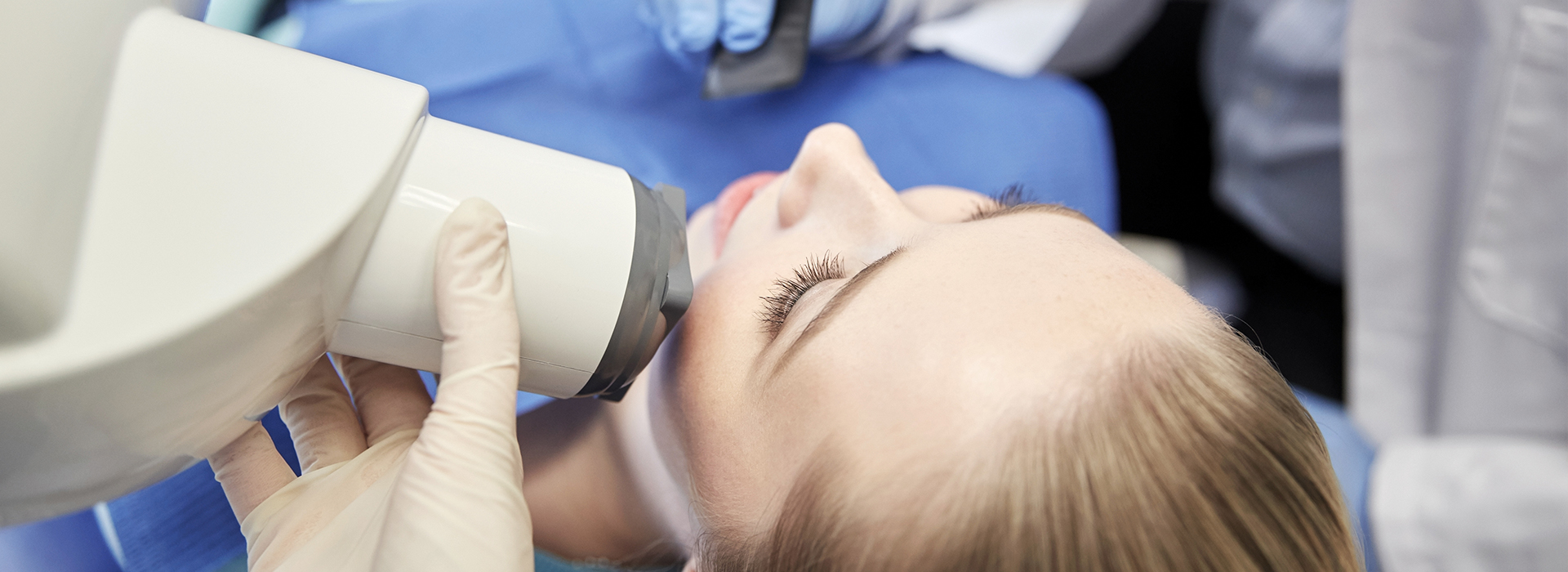
Digital radiography replaces traditional film with electronic sensors and computer processing to capture dental images. Instead of a chemical-developing workflow, an image is recorded on a sensor and translated immediately into a high-resolution file. The result is a clearer, more flexible picture of teeth, roots, and supporting bone that can be examined in real time during your appointment.
Because the image appears instantly on a chairside monitor, clinicians can evaluate findings on the spot and explain what they see to patients more effectively. This immediacy reduces the need to wait for film to develop, speeds clinical decision-making, and helps patients understand proposed treatments through on-screen visualization.
Beyond convenience, the digital process streamlines documentation. Images are saved to a secure electronic record tied to each patient’s chart, simplifying long-term tracking and comparisons. This is especially helpful when monitoring changes over time or coordinating care with other dental professionals.
One of the primary advantages of digital radiography is a reduction in the radiation dose required to produce a diagnostic image. Modern sensors are more sensitive than film, so clinicians can capture usable images with lower exposure levels while still maintaining diagnostic quality. In practice, this contributes to a safer imaging experience for patients of all ages.
Digital imaging also reduces the need for repeat exposures. Software tools allow technicians to adjust contrast, brightness, and cropping to enhance an image without taking another x‑ray. Fewer retakes mean less cumulative exposure and a smoother, quicker visit for patients.
From an environmental perspective, going digital eliminates the chemical processing and paper-based workflows associated with film. There are no developer solutions, fixer chemicals, or physical prints that need disposal, which simplifies clinic operations and reduces ecological impact.
Digital sensors produce images with strong resolution and consistent quality, giving dentists reliable visual information for diagnosing cavities, assessing bone levels, and evaluating root structures. Enhanced image clarity helps clinicians detect subtle changes earlier, which can lead to more conservative and effective treatment choices.
Integrated software provides measurement and annotation tools that support precise treatment planning. For example, clinicians can measure the distance between anatomical landmarks or magnify an area to inspect fine detail without degrading image quality. These capabilities are essential for procedures that require careful spatial planning, such as implant placement or root canal therapy.
Because files are stored digitally, clinicians can compare current images with prior studies side by side. This chronological view makes it easier to spot gradual changes in tooth structure, bone density, or restorative work, supporting better long-term monitoring and more informed follow-up care.
Digital images are also adaptable for multidisciplinary collaboration. Specialists can review the same files remotely, and images can be exported in standard formats for use in treatment planning software or second opinions when needed, improving the overall quality and coordination of care.
Digital radiography speeds routine workflows from capture to recordkeeping. Once an image is taken, it is automatically attached to the patient’s electronic chart and becomes available to the entire care team. This reduces administrative steps and minimizes the chance that an image will be misplaced or misfiled.
Instant access supports more efficient visits: clinicians can confirm findings, show images to patients, and document treatment recommendations in one session. That same access helps the team triage urgent issues more rapidly and prepare appropriate instruments or referrals while the patient is still in the office.
Secure digital storage also simplifies sharing with outside providers when coordination is required. With appropriate patient consent, files can be transmitted to specialists for review without the delay or logistics of shipping physical films. This timeliness can improve patient experiences and shorten the interval between diagnosis and definitive care.
At Value Dental Center, our clinical workflow leverages digital imaging to reduce appointment time and enhance communication between clinicians and patients, so care is both efficient and easy to follow.
Undergoing a digital dental x‑ray is quick and straightforward. A small sensor is positioned inside the mouth or an external device is placed near the jaw for panoramic imaging; the clinician will help you get into a comfortable position and use supportive bite blocks or cushions as needed. The actual exposure lasts only a fraction of a second in most cases.
Because digital sensors are slimmer and more flexible than traditional film holders, many patients find the process more comfortable. Protective measures—such as a lead apron when appropriate—are used to reduce exposure to surrounding tissues, and the imaging team follows standardized protocols to ensure consistent safety practices.
After images are captured, the clinician will review them on a monitor and explain what they show. This review typically includes pointing out areas of decay, bone loss, or existing restorative work, as well as discussing next steps for treatment or monitoring. Patients appreciate seeing the same images the clinician uses to make recommendations, which supports clear, informed decision-making.
Digital radiography is a dependable, patient-focused tool that enhances safety, speeds clinical workflows, and improves diagnostic clarity. If you’d like to learn more about how digital imaging is used in our office or how it can support your dental care, contact Value Dental Center for more information.
Digital radiography is a dental imaging method that uses electronic sensors and computer software to capture, view and store X-ray images. Instead of film, a small sensor inside the mouth or an external digital unit captures the image and the data is transferred directly to a computer where it can be enhanced and reviewed. This technology streamlines the imaging process and supports faster diagnosis compared with traditional film systems.
Digital images can be adjusted for contrast and magnification to help clinicians evaluate tooth structure and bone levels more precisely. Because the files are electronic, they can be stored in a secure patient record and shared with other providers when coordination of care is needed. The approach reduces the need for chemical processing and physical storage, making it a more efficient clinical workflow.
Digital radiography replaces photographic film with electronic sensors that are more sensitive to X-ray photons, so images are produced with less radiation and appear on a monitor instantly. Unlike film, digital images can be enhanced, measured and compared side-by-side using software tools, which helps clinicians identify subtle changes over time. Film requires chemical development, physical storage and more time between exposure and review, while digital systems eliminate those steps.
Digital files can be duplicated without degradation, transmitted electronically to specialists and integrated into an electronic health record for long-term tracking. The immediate feedback and image manipulation options improve diagnostic efficiency and patient communication. Environmental advantages include eliminating processing chemicals and reducing paper or film waste.
Digital sensors are more sensitive than traditional film and therefore require lower X-ray doses to produce a diagnostic-quality image. Modern digital protocols also combine optimized exposure settings, beam collimation and high-speed sensors to keep exposure as low as reasonably achievable, following standard radiation-safety practices. Clinicians select the smallest field of view and the fewest images necessary to answer the clinical question.
Additional protective measures such as lead aprons and thyroid collars are used when appropriate to shield sensitive tissues. Children and other radiation-sensitive patients receive special consideration with adjusted exposure settings and reduced image frequency. If you have specific concerns about exposure, discuss them with your dental team so they can explain the safeguards used during your visit.
During a digital radiography appointment, the dental team will position a small sensor or digital plate in your mouth while the X-ray unit is aligned externally to capture the image. The sensor is connected to a computer, so the image appears on a monitor almost immediately and the clinician can confirm whether additional views are necessary. The process is quick and typically causes minimal discomfort beyond holding the sensor in position for a few moments.
Depending on the clinical need, your appointment may include bitewing, periapical or panoramic images and each type serves a different diagnostic purpose. Protective equipment such as a lead apron may be used, especially for children or patients with heightened sensitivity. The clinician will review the images with you and explain any findings or next steps during the same visit.
Digital radiography is considered safe for children when exposures are justified and minimized, because modern sensors allow for much lower radiation doses than older film systems. Pediatric patients receive tailored exposure settings and positioning aids to reduce retakes and improve comfort, and clinicians follow recommended guidelines for frequency and necessity of images. Parents are encouraged to discuss any specific concerns or medical history that might affect imaging decisions.
For pregnant patients, dental X-rays are generally deferred when possible during the first trimester unless the imaging is essential for diagnosing an urgent condition. When imaging is required, extra protective measures such as a lead apron and thyroid shield are used to minimize exposure, and the dental team will document the justification for the exam. Always inform your dental provider if you are pregnant so they can adapt the plan and discuss alternatives.
Digital dental images are stored electronically in secure patient records, typically within a practice management system or a picture archiving and communication system (PACS). These systems use encryption, user authentication and role-based access controls to limit who can view or modify patient images. Regular backups and disaster-recovery processes are implemented to preserve data integrity and continuity of care.
Images can be shared with other providers or specialists when you authorize the transfer, which speeds consultations and collaborative treatment planning. The practice follows applicable privacy laws and professional standards to protect patient information and only releases records according to legal and ethical guidelines. If you have questions about how your images will be used or stored, ask the dental team for a clear explanation of their data-protection procedures.
Digital radiography is a powerful diagnostic tool for detecting cavities between teeth, assessing bone levels around teeth, identifying root infections and evaluating other hard-tissue conditions. However, it does not show every condition, and some problems such as early surface decay, soft-tissue lesions or certain types of fractures may require additional clinical evaluation or alternative imaging. The clinician combines radiographic findings with a visual exam, patient history and other tests to form a complete diagnosis.
For advanced or three-dimensional assessments, such as implant planning or complex pathology, cone-beam computed tomography (CBCT) or other imaging modalities may be recommended in addition to standard digital X-rays. Your dental provider will explain the strengths and limitations of each imaging option and recommend the most appropriate approach for your specific needs. Imaging decisions are driven by the clinical question and the goal of obtaining the information necessary for safe, effective care.
Digital X-rays allow dentists to view, measure and annotate images during an appointment, which supports precise treatment planning and clearer explanations for patients. The ability to enhance contrast, zoom in on areas of concern and compare current images with past records helps clinicians track disease progression and evaluate treatment outcomes. Digital files can also be sent quickly to laboratories or specialists to coordinate care without delay.
Using on-screen images during consultations improves patient understanding and engagement by showing the exact findings being discussed. This visual approach helps patients make informed decisions about recommended treatments and follow-up care. Sharing images electronically with collaborating providers also reduces the need for duplicate exposures and supports continuity of care.
One of the advantages of digital radiography is near-instantaneous results; images typically appear on the clinician's monitor within seconds of capture. This immediate availability allows the dental team to review findings and discuss them with you during the same visit, which expedites diagnosis and planning. If you want a copy of your images, many practices can provide printed images or digital files upon request, consistent with privacy and record-release policies.
Viewing images together with the clinician helps you understand the condition of your teeth and supporting structures and allows for real-time questions about treatment options. Digital storage also makes it easy to retrieve past images for comparison at future visits. If you have trouble interpreting what you see, ask the dental team to walk you through the image and explain relevant markers or measurements.
Routine maintenance, calibration and quality-assurance checks are essential to ensure consistent image quality and safe operation of digital radiography equipment. The practice follows manufacturer recommendations and industry standards for sensor care, X-ray unit performance testing and software updates, and staff receive training on proper positioning and exposure techniques. Disinfection protocols for intraoral sensors and barriers are used between patients to maintain infection-control standards.
At Value Dental Center, the clinical team adheres to these best practices and documents equipment checks as part of their quality program. Regular audits and continuing education help keep imaging processes current with evolving safety guidelines and technological advances. If you have questions about the practice's equipment maintenance or safety procedures, the team can provide specific information during your appointment.
Quick Links