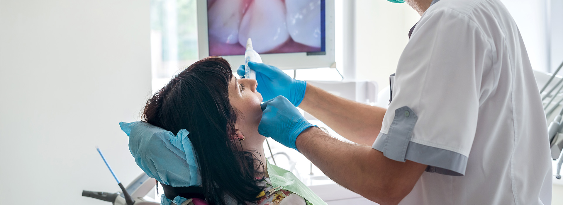
Modern dental care blends skilled clinical judgment with technologies that make diagnosis clearer and treatment more predictable. The intraoral camera is one such tool: a compact, pen-sized imaging device that provides detailed, full-color views of the teeth and soft tissues and displays them on a monitor in real time. Used routinely during exams and treatment discussions, this device helps clinicians and patients observe the mouth at a level of detail that’s difficult to achieve with unaided vision alone. At Value Dental Center, we use intraoral imaging to support clear communication and precise clinical decision-making.
Intraoral cameras capture high-resolution images of individual teeth, restorations, gum margins, and areas of concern such as cracks, wear facets, and early decay. Unlike traditional mirror-based inspection, the camera can be positioned to show tight or hard-to-see spaces from multiple angles while keeping the patient comfortable. The resulting images are full-color and magnified, making subtle changes in texture, color, and contour easier to identify.
Because the camera produces photographic-quality images, clinicians can compare current views with past images to monitor progression or healing over time. This objective visual record supports more accurate charting and helps the care team detect problems earlier, often before symptoms appear. The technology is particularly useful for spotting small fractures, marginal gaps around fillings, or the beginning stages of enamel demineralization.
Images are not limited to static photographs; video capture is also possible on many systems, allowing clinicians to document dynamic issues such as a bite relationship or movement of a restoration. These visual records become part of the patient’s clinical record, supporting continuity of care and more informed treatment planning between general dentists and specialists when collaboration is needed.
An intraoral camera transforms abstract clinical findings into clear visual information that patients can see and understand. Rather than relying on verbal descriptions or X-ray interpretations alone, clinicians can show live images on a screen and walk patients through what they are seeing. This visual approach encourages questions and helps patients make informed decisions about recommended treatments.
When patients view their own oral condition, they gain a more accurate understanding of the relative urgency and goals of care. Clear images demystify common issues—such as the early signs of decay or worn restoration margins—and reduce uncertainty that can otherwise hinder acceptance of necessary treatment. This shared visual reference fosters a collaborative relationship between clinician and patient.
Intraoral images are also valuable educational tools. Care teams can annotate images or point to specific landmarks during explanations, reinforcing hygiene instruction or demonstrating how disease progresses without intervention. Patients leaving the office with a visual memory of the discussion are more likely to follow preventive guidance and adhere to recommended recall schedules.
From a clinical perspective, intraoral cameras contribute to more precise diagnoses. Fine details that are easily missed during a cursory exam become visible under magnification, allowing earlier detection of problems that influence treatment choice—from minimally invasive repairs to full restorations. This early detection can preserve more natural tooth structure and simplify future care.
For restorative treatments, intraoral imaging helps evaluate the fit and margins of crowns, bridges, and fillings. It aids in documenting the initial condition before treatment, verifying work after placement, and monitoring how restorations perform over time. In cases involving multiple providers, shared images create a clear and consistent record that supports coordinated care and predictable outcomes.
Periodontal assessment also benefits from intraoral visualization. Images of gum margins, recession, and plaque accumulation help the team assess periodontal health and track responses to therapy. The objective nature of the photographs reduces ambiguity in treatment planning and supports evidence-based decision-making.
Capturing intraoral images is a straightforward process that integrates into routine appointments. After a brief explanation, the clinician uses the camera to scan target areas while the images appear instantly on a monitor. Many systems allow stills and short videos to be saved directly into the patient’s digital chart. This process typically adds only minutes to an exam but provides substantial value in documentation.
Once stored, images serve multiple clinical purposes: they become part of the permanent record for comparison during follow-up visits, they can be shared securely with specialists or a dental laboratory when coordinated care or prosthetic fabrication is needed, and they support claims documentation when clinical justification is required. Properly managed digital images enhance transparency and continuity across all phases of care.
Security and privacy are essential when handling digital images. Practices that use intraoral cameras follow standard protocols to protect patient information, ensuring that stored images remain part of the confidential health record and are accessed only by authorized team members as part of treatment delivery.
Patients typically find intraoral imaging quick and noninvasive. The device resembles a small wand and is gently introduced into the mouth while the clinician asks the patient to open or reposition as needed for the best view. The camera’s LED lighting ensures images are bright and clear; most patients experience no discomfort beyond normal exam maneuvers.
Because images appear on a screen in real time, patients can follow along as the clinician explains findings. This immediate visual feedback helps clarify the nature of any concern and the reasons behind recommended next steps. For anxious patients, seeing exactly what the clinician sees often reduces uncertainty and increases confidence in the proposed care plan.
After the exam, key images are saved to the chart for future reference, and the clinician will summarize the findings and recommended options. Patients who want to review what was shown can request additional explanation or be directed to educational resources the practice provides for further reading.
Intraoral cameras have become an essential component of modern dental practice because they bring clarity, documentation, and patient engagement to routine care. By making small problems visible early and helping patients understand their oral health, this technology supports better outcomes and more collaborative treatment decisions. If you would like to learn more about how intraoral imaging is used in our office or have questions about your next visit, please contact us for more information.
An intraoral camera is a pen-sized, high-resolution camera that captures detailed color images inside the mouth and displays them in real time on a computer monitor. The device provides magnified views of teeth, gums, and other oral tissues that are difficult to see with the naked eye. Images can be captured instantly for review, discussion, and documentation.
Clinicians use the intraoral camera to examine surfaces, identify signs of wear or damage, and record findings in the patient record. At Value Dental Center we rely on this technology to improve diagnostic clarity and involve patients directly in their care decisions.
The intraoral camera gives clinicians a close-up, high-definition view of tooth surfaces and soft tissues that enhances the detection of fractures, cracks, staining, and plaque accumulation. This magnified perspective helps identify areas that require further evaluation and supports more precise treatment recommendations. Because images are recorded, clinicians can compare changes over time to track progression or healing.
Captured images integrate with digital records and treatment planning tools to guide restorative work, periodontal care, and preventive strategies. When combined with radiographs and clinical exams, intraoral photos contribute to a comprehensive diagnostic picture and more predictable outcomes.
Yes, intraoral cameras are noninvasive and safe for routine clinical use. They do not emit ionizing radiation and simply record visible light images of the oral cavity. Disposable sheaths or barriers are used for infection control and the device is cleaned according to established sterilization protocols to protect patients.
Operators are trained to handle the camera gently and position it to minimize discomfort while obtaining clear images. The process is typically brief and well tolerated by most patients, including those with dental anxiety.
An intraoral camera lets patients see what the dentist sees by displaying live images on a screen, which makes explanations of conditions and treatment options more concrete and understandable. Visual evidence helps patients recognize the location and extent of problems such as cavities, gum inflammation, or worn restorations. This transparency supports informed consent and encourages collaboration on care decisions.
When clinicians review images with patients, it becomes easier to prioritize treatments and demonstrate the expected results of preventive or restorative steps. Value Dental Center uses intraoral imaging as part of its educational approach to help patients feel informed and engaged in their oral health journey.
An intraoral camera appointment typically involves brief image capture of specific teeth or areas of concern while you sit comfortably in the dental chair. The clinician will gently position the camera inside the mouth and angle it to show the targeted surfaces; you may be asked to open or move slightly to improve visibility. Most captures take only a few seconds per image and are painless.
After images are taken, the clinician will review them on-screen with you and explain any findings or recommended next steps. Because images are saved, they can be used for follow-up comparisons or to document the condition before and after treatment.
Images from the intraoral camera are stored digitally within your secure dental record alongside clinical notes and radiographs. These images become part of the permanent chart and can be retrieved for future visits to monitor changes or evaluate the effectiveness of treatment. Digital storage also facilitates clear documentation of conditions discussed during an appointment.
Stored images are used to coordinate care with specialists, guide laboratory work for restorations, and support communication within the clinical team. Access and sharing follow applicable privacy and data-protection practices to safeguard patient information.
Intraoral cameras are effective at revealing visible surface changes such as staining, cracks, chips, and surface demineralization that may indicate early decay. They are particularly useful for documenting discoloration and small defects that are hard to see unaided. However, some cavities that form between teeth or below the surface are best detected with radiographs or other diagnostic tools.
Because of these limitations, intraoral imaging is used alongside dental examinations and X-rays to provide a full assessment. The combined approach improves early detection and helps determine whether preventive care, monitoring, or restorative treatment is appropriate.
An intraoral camera captures surface-level color images showing texture, color, and soft-tissue detail, while dental X-rays provide information about structures beneath the surface such as tooth roots, bone levels, and interproximal decay. Each modality reveals different aspects of oral health and neither fully replaces the other. Cameras excel at patient-facing visuals and surface documentation; radiographs are essential for diagnosing hidden problems.
Dentists commonly use both intraoral imaging and radiography in tandem to form a complete diagnostic picture. This complementary use enables safer, more accurate treatment planning by combining visual surface data with internal structural information.
Yes, captured images can be exported and shared with dental specialists, laboratories, or other authorized parties to support referrals and collaborative care. High-quality photos help specialists evaluate a case remotely and laboratories use images to match shade, shape, and margin details for restorations. Sharing is performed with patient consent and follows privacy and record-transfer protocols.
Using intraoral images during referrals and lab prescriptions improves communication, reduces ambiguity, and can accelerate the treatment process. Clear visual documentation helps ensure that the receiving clinician or technician understands the clinical situation and desired outcomes.
Intraoral images allow clinicians and patients to examine details such as tooth alignment, staining, worn edges, and existing restorations that influence cosmetic or restorative decisions. Photographs serve as a visual baseline for planning veneers, crowns, whitening, and other procedures, and they assist in communicating goals with dental laboratories. The ability to capture and review multiple angles improves accuracy when designing restorations or cosmetic enhancements.
During treatment planning, images can be annotated or compared to reference cases to clarify expectations and procedural steps. This visual documentation supports predictable, esthetic outcomes and helps the clinical team coordinate technical specifications with the dental lab.
Quick Links